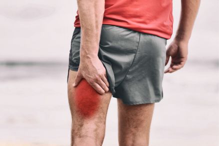
Tendinitis refers to inflammation and irritation of a tendon, which is the thick fibrous cord that attaches muscle to bone. Tendons are comprised of small fibers of collagen that gradually merge together into a larger tendon unit. Inflammation makes these fibers slide unevenly against each other instead of gliding smoothly. This causes pain and discomfort, especially with movement or use of the affected joint. Tendinitis can occur in any tendons, but most commonly affects the shoulders, elbows, wrists, knees, and heels.
Causes of Tendinitis
There are several factors that can contribute to developing tendinitis:
- Overuse – Repeated motions or overexertion of a tendon through sports, work, or other activities can lead to microtears in the collagen fibers. For example, baseball pitching, swimming laps, running long distances, and repetitive tasks like painting or typing all involve the overuse of certain tendons. The damage adds up over time until symptoms of irritation and inflammation arise.
- Sudden injury – Direct trauma like falls or collisions can cause injury to a tendon. Even a strong sudden contraction against resistance can tear tendon fibers.
- Anatomic problems – Issues with musculoskeletal alignment or structure can increase strain on certain tendons and predispose someone to tendinitis. This includes having flat feet, knocked knees, bowed legs, or an imbalance in leg length.
- Age – As people get older, tendons become less flexible and elastic. This makes them more vulnerable to small tears and inflammation, especially if exercise or activity levels are suddenly increased.
- Underlying diseases – Some health conditions are associated with a higher risk of tendinitis. These include rheumatoid arthritis, gout, lupus, and diabetes.
- Medications – Certain antibiotics like fluoroquinolones have been linked to tendon damage and a higher incidence of tendinitis. Long-term oral or injected steroid use can also sometimes weaken tendons and make them more prone to inflammation.
- Poor circulation – Reduced blood supply to tendons likely impairs their ability to heal after microinjuries. This can allow tendinitis to develop more easily. Smoking impairs circulation and has been associated with higher risk.
Any of these factors result in small tears in the tendon fibers, causing the body to flood the area with inflammatory chemicals and cells meant to start the healing process. This leads to irritation, swelling, tenderness, and pain around the affected tendon. If the injury is minor, rest and modifying activities will allow proper healing and resolution of symptoms. However, if there is repeated overuse or aggravation without adequate rest, chronic tendinitis can occur. This leads to long-term thickening and weakness of the tendon.
Common Sites of Tendinitis
There are certain tendons that are more prone to becoming inflamed with overuse injuries. Common locations include:
Shoulder Tendinitis
Two of the most commonly injured groups of tendons in the shoulder are the rotator cuff and biceps tendons. The rotator cuff is comprised of four muscles around the shoulder blade that control intricate motions of the shoulder joint. Repeated overhead motions like serving a tennis ball, pitching a baseball, or painting a ceiling can lead to rotator cuff tendinitis. This affects the tendons of the supraspinatus, infraspinatus, subscapularis, and teres minor muscles, causing pain at the top or front of the shoulder that worsens with reaching or overhead movements.
Biceps tendinitis causes irritation and inflammation of the long head of the biceps tendon. This can happen from repetitive heavy lifting or flexion against resistance. Pain is focused at the front of the shoulder and may radiate down the upper arm. Sports like swimming, tennis, and baseball can lead to the overuse of biceps tendinitis. Another type of shoulder tendinitis is adhesive capsulitis, which involves inflammation of the ligaments of the shoulder capsule rather than a specific tendon.
Elbow Tendinitis
Lateral and medial epicondylitis, also known as tennis elbow and golfer’s elbow, are two common forms of tendinitis affecting the elbow. Tennis elbow involves the tendons attaching the forearm extensor muscles to the lateral epicondyle of the humerus. It is commonly caused by repetitive wrist extension and forearm rotation, as seen with backhand strokes in tennis or other racket sports. Golfer’s elbow affects the tendons attaching forearm flexor muscles to the medial epicondyle. It is often triggered by repetitive wrist flexion and forearm rotation involved in the golf swing and other sports relying heavily on the wrist. Jobs involving repetitive gripping, wrist flexion, and forearm rotation like carpentry and painting can also lead to medial elbow tendinitis.
Wrist Tendinitis
The most frequently injured tendons in the wrist are on the thumb side and include the digital extensor tendons passing under the extensor retinaculum. De Quervain’s tenosynovitis involves irritation where the tendons for the thumb extensor pass through the first extensor compartment. It is often triggered by repetitive grasping and twisting motions of the wrist and thumb. Flexor tendinitis most commonly affects the superficial and deep digital flexor tendons along the palm side of the forearm and under the flexor retinaculum of the wrist. It can result from frequent repetitive flexion of the fingers and wrist. Frequent typing is often implicated in flexor tendinitis of the wrist. Carpal tunnel syndrome is sometimes mislabeled as wrist tendinitis, but it actually involves compression of the median nerve rather than inflammation of the wrist tendons. However, flexor tendon swelling can worsen carpal tunnel syndrome.
Knee Tendinitis
Some of the largest and most heavily used tendons pass around the knee joint. The quadriceps tendon connects the quadriceps muscles of the thigh to the patella and then continues down to the tibia as the patellar tendon. Patellar tendinitis, also called jumper’s knee, involves inflammation of the patellar tendon as it inserts into the tibia below the kneecap. Pes anserine tendinitis affects the conjoined tendons of the hamstring muscles where they attach to the tibia on the inside of the knee. Both types of knee tendinitis result from frequent running, jumping, and impact on a bent knee involved in many sports. The iliotibial band running along the outside of the thigh can also become inflamed with repetitive knee flexion.
Achilles Tendinitis
The large Achilles tendon attaches the gastrocnemius and soleus calf muscles to the heel bone. As the strongest tendon in the body, it experiences significant forces during running, jumping, and pushing off the ball of the foot. This makes it prone to overuse tendinitis in sports like track, basketball, tennis, and soccer. Achilles tendinitis causes pain and stiffness focused in the back of the ankle which is often worse with activity. Bone spurs can form over time at the insertion of the tendon. Minor calf tears are also commonly misdiagnosed as Achilles tendinitis.
Symptoms of Tendinitis
Tendinitis causes characteristic symptoms near joints like:
- Pain – typically worse at the start of an activity and easing somewhat once tendons warm up and limber up with use. It returns once the activity stops.
- Tenderness – Focused at the point where inflamed tendons insert into bone, usually just outside a joint capsule. Pressure on the area is uncomfortable.
- Stiffness – More pronounced after periods of inactivity and improving with movement. Generalized stiffness around the joint is common.
- Swelling – Mild inflammation noticeable a few hours after repetitive overuse of a tendon. Visible swelling is less common.
- Warmth – Increased warmth around the inflamed tendon compared to surrounding areas, though often only mildly detectable.
- Reduced range of motion – Active and passive joint movement may be restricted due to discomfort and swelling.
As tendinitis becomes more severe or chronic without proper rest and treatment, symptoms often worsen and become more persistent:
- Pain interferes with daily tasks and sleep
- Stiffness lasts for hours instead of minutes after activity
- Joint strength and mobility progressively decline
- Noticeable swelling lasts for days instead of hours
- Crepitus – cracking or crunching sound when moving the joint
- Locking – brief inability to fully straighten a joint
Diagnosing Tendinitis
To diagnose tendinitis, a physician will start with a medical history looking at activity, any trauma, other medical conditions, and prior injuries. Description of the location, nature, timing, and aggravating factors of the pain can help identify the affected tendon. Physical examination will focus on inspection, range of motion testing, palpation for tenderness, and provocative maneuvers to reproduce symptoms.
With the elbow, wrist, or knee, palpating the joint line will often pinpoint tenderness at tendon insertions. Resisted movements can differentiate tendinitis from muscle or joint issues. Shoulder and Achilles tendinitis can be harder to distinguish from other injuries without imaging. Provocative tests like the arc sign and chair push-up test for shoulder tendinitis can aid in diagnosis.
If the clinical exam is equivocal, imaging studies may be ordered to confirm tendinitis or rule out other problems. Useful imaging tests include:
- X-rays – Best for detecting bony changes like calcification in chronic tendinitis. Not as helpful for soft tissue inflammation.
- MRI – Provides clear images of swelling, fluid, and damage in and around tendons and joints. Extremely useful for diagnosing tendinitis.
- Ultrasound – Allows dynamic visual evaluation of tendons in motion. Can show swelling, calcifications, and partial tears. Very useful for shoulder tendinitis diagnosis.
- CT scan – Provides excellent visualization of bony deformities that could irritate tendons. Less useful for soft tissue evaluation.
Treatment for Tendinitis
Treatment focuses on relieving pain and inflammation while promoting healing of the tendon tissue. A combination of approaches is often used including:
- Rest – Avoiding activities that aggravate the inflamed tendon prevents further injury and encourages natural healing. splints, slings, or crutches may be used to immobilize joints.
- Ice – Applying ice for 10-15 minutes several times a day constricts local blood vessels, reducing swelling and inflammation. Cold therapy is most helpful in early symptomatic stages.
- Compression – Snug compression bandages or sleeves help limit swelling and provide support. Used in combination with ice and elevation.
- Elevation – Keeping the sore joint elevated above the level of the heart utilizes gravity to drain fluid from the area and minimize swelling.
- NSAIDs – Anti-inflammatory drugs like ibuprofen block the production of inflammatory prostaglandins, relieving pain and swelling. Topical diclofenac gel can directly treat affected tendons.
- Injections – Cortisone shots block inflammation pathways and reduce swelling/irritation in and around the tendon. Platelet-rich plasma contains growth factors to help repair tendon tissue.
- Stretching – Gentle daily stretching keeps joints mobile and flexible while avoiding further straining the inflamed tendon. Can restore normal motion once acute inflammation resolves.
- Eccentric exercises – Controlled progressive loading through eccentric contractions helps rebuild collagen strength and flexibility in the damaged tendon.
- Massage – Gentle transverse friction massage perpendicular to the tendon can break up adhesions and reduce thickening in chronic tendinitis.
- Heating pads – Applying localized heat before activities or exercise can relax muscles and stimulate blood flow around chronically inflamed tendons.
- TENS – Applying transcutaneous electrical nerve stimulation can provide symptomatic pain relief at the tendinitis site.
- Night splints – Immobilizing braces worn during sleep stretch tissues and prevent morning stiffness in wrists and ankles.
- Shockwave therapy – Pulsed acoustic sound waves seem to stimulate healing responses in damaged tendon tissue, though mechanisms are not fully understood.
- Surgery – Rarely required as a last resort for chronic recalcitrant cases involving tendon tears or bone spurs irritating tendons. Options range from debridement to resection and repair.
Most cases of acute tendinitis resolve within a few weeks with sufficient rest coupled with therapies to control inflammation and accelerate tendon healing. However, neglected or recurrent tendinitis can become chronic, requiring more intensive rehabilitation and treatments to restore normal tendon gliding function. Prevention through regular stretching and gradual conditioning along with avoiding overexertion or overuse is key to reducing future tendinitis flares. Proper treatment combined with activity modification helps the majority of tendinitis patients achieve full resolution of symptoms and return to normal activity.
Risk Factors for Developing Tendinitis
Certain individuals are at increased risk for developing tendinitis based on their occupation, activities, health status, and body mechanics. Risk factors include:
- Sports with repetitive arm motions – Examples are tennis, swimming, baseball, cross-country skiing, and track/running. Overhead arm motions like serving or freestyle swimming are especially risky.
- Sports with jumping – Basketball, volleyball, track & field, and soccer involve repetitive impact and explosive push-off that can strain lower extremity tendons like the patellar and Achilles.
- Racket sports – Tennis, racquetball, squash, and badminton involve forceful gripping and wrist motions that strain the extensor and flexor forearm tendons.
- Forceful physical jobs – Construction, manufacturing, painting, carpentry, and plumbing often require repetitive heavy gripping, lifting, and awkward positions leading to tendinitis.
- Office jobs with repetitive motions – Extensive typing, computer mouse work, and writing increase the risk for wrist/hand tendinitis. Prolonged sitting can also contribute to poor posture.
- Age over 30 – The elasticity and cell repair ability of tendons decreases with aging, making them less able to recover from microtears.
- Deconditioning – Weak or inflexible muscles attached to a tendon pull harder on the tendon fibers instead of absorbing force themselves. Tight hamstrings are correlated.
- Obesity – Excess body weight puts greater tension on weight-bearing tendons like the patellar and Achilles tendons.
- Anatomic misalignments – Having flat feet, bowed legs, knock knees, or difference in leg length alters mechanics in a way that increases tendon strain.
- Systemic diseases – Rheumatoid arthritis, lupus, gout, and diabetes are linked to higher incidence of tendinopathy and tendinitis.
- Certain medications – Antibiotics like Cipro or prednisone have adverse impacts on tendon integrity and elasticity.
- Previous tendon injuries – Once a tendon is damaged, it is more susceptible to re-injury and slower to heal with future bouts of tendinitis.
Knowing these predisposing factors can help target preventive strategies, conditioning programs, and early conservative treatment to minimize tendinitis susceptibility. At-risk individuals should take extra precautions like pacing activities gradually, using proper technique, maintaining strength and flexibility, avoiding excessive loading, and managing underlying medical conditions. This comprehensive approach is key to reducing the likelihood of initial and recurrent tendinitis.
Preventing Tendinitis
Many cases of tendinitis can be avoided by taking preventive measures to decrease tendon strain, improve tendon conditioning, and avoid aggravating activities. Recommended injury prevention strategies include:
- Warming up adequately before sports or vigorous activity – Gradually increasing the intensity and duration of warm-up activities enhances tendon extensibility, elasticity, and blood flow.
- Stretching regularly – Gentle daily stretching maintains flexibility and range of motion, reducing the risk of overstretching tendons. Focus on calf, thigh, forearm, and shoulder stretches.
- Strengthening exercises – Building strength in muscles attached to vulnerable tendons helps absorb forces rather than overloading tendons. Target legs, shoulders, and wrists.
- Cross-training – Varying sports and workouts avoid overuse of the same movements and tendons.
- Improving technique – Ensure proper form and mechanics during repetitive motions at work or play to avoid putting excess strain on joints.
- Using ergonomic equipment – Chairs, keyboards, tools, and sports equipment tailored to the user protects tendons from excessive strain.
- Taking frequent breaks – Interspersing repetitive tasks with short rest periods allows tendons time to recover between bouts of activity.
- Modifying or avoiding pain-provoking activities – Stop motions that acutely aggravate tendinitis symptoms until fully healed.
- Wearing braces or supports – Compression sleeves decrease tendon strain and irritation during suspect activities.
- Gradually increasing activity levels – Slowly building up duration, intensity, and frequency puts less sudden stress on tendons.
- Applying ice after strenuous activity – Cold therapy constricts vessels and decreases early inflammation and swelling to prevent overuse irritation.
By being proactive and minimizing tendon overexertion and strain, most people can avoid episodes of painful tendinitis throughout the musculoskeletal system. A combination of tailored strengthening exercises, sufficient rest periods, proper technique, supportive equipment, and gradually ramping up new activities allows tendons time to adapt without incurring damage. Remaining vigilant for early signs of tendon irritation and modifying activities appropriately helps stop acute tendinitis from developing into a chronic problem. With some diligence about prevention, tendinitis can usually be avoided.
Chronic Tendinitis Treatment
In some cases, tendinitis is not adequately treated or activities continue that perpetuate microtears and inflammation in the tendon. This can lead to a chronic tendinopathy that is much more resistant to conservative rehabilitation measures. Key steps in treating chronic recalcitrant tendinitis include:
- Extended activity cessation – Refraining from implicated activities for 2-6 weeks allows inflammation to resolve and prevents further repetitive injury.
- Aggressive stretching – Regularly apply gentle sustained stretches to the tendon and attached muscle to restore normal excursion.
- Eccentric exercises – Controlled progressive loading through eccentric muscle contractions specifically rebuild tendon strength and elasticity.
- Deep friction cross-fiber massage – Mobilizes thickened tendon adhesions and scar tissue to improve flexibility.
- Glyceryl trinitrate patches – Nitric oxide promotes collagen synthesis and has shown improvement in tendinosis and tears.
- Extracorporeal shockwave therapy – Acoustic sound waves applied to the tendon area seem to stimulate the healing and regeneration of chronically damaged tissue.
- Platelet-rich plasma injections – Injecting concentrated growth factors from the patient’s own blood into the tendon encourages healing and repair.
- Hyaluronic acid injections – Viscous injections may help lubricate and loosen adhesions between tendon fibers.
- Tenotomy – Surgically releasing tension in the tendon may be considered if hypercontracted and resistant to conservative measures.
- Tendon debridement – Removing degraded tissue or bony spurs rubbing on the tendon.
- Tendon graft – In cases of severe chronic tendinosis, surgically implanting a donor tendon graft can functionally replace the damaged tendon.
- Orthotics – Custom arch supports and braces correct any biomechanical issues contributing to tendon strain and irritation.
- Alternative therapies – Treatments like acupuncture, massage, ultrasound, and magnet therapy may provide symptomatic relief in some refractory cases.
The key to treating chronic tendinitis is starting an aggressive rehabilitation protocol focusing on flexibility, eccentric strengthening, and eliminating irritation from impinging motions or arthritis. Realistic activity modification is necessary to prevent ongoing tendon overuse and allow proper healing. If significant functional impairment persists despite exhaustive conservative efforts, surgical options like debridement or grafting may be warranted. A personalized treatment plan tailored to the individual’s daily activities, functional demands, and prior treatment response offers the best prospects for eventually overcoming chronic recalcitrant tendinitis.
Achilles Tendinitis
The Achilles tendon attaches the calf muscles to the back of the heel bone, allowing plantarflexion of the ankle and foot. As the strongest and thickest tendon in the body, it is subjected to very high tensile loads during running, jumping, and push-off motions. These explosive forces concentrated on the Achilles tendon make it vulnerable to overuse injuries and tendinitis. Achilles tendinitis classically causes pain and stiffness at the back of the heel that is aggravated by athletic activity. It can involve the mid-portion of the tendon or its insertion into the calcaneus bone.
Contributing factors for Achilles tendinitis include tight calf muscles reducing flexibility, sudden increases in training intensity or duration, improper foot biomechanics like overpronation, and wearing high-heeled shoes which increase strain on the tendon. It is particularly common in runners and athletes involved in sports that require frequent jumping like basketball, volleyball, tennis, and soccer. Middle-aged non-athletes can also develop Achilles tendinitis just from increasing walking duration or intensity too abruptly.
Symptoms consist of pain and stiffness focused at the back of the ankle near the heel. It is usually worse with the first steps after getting up in the morning or at the beginning of athletic activity. Mild swelling may be present. Moving the ankle feels stiff and achy at first but tends to loosen up as the tendon warms up. In chronic cases, bone spurs can form near the tendon insertion.
To treat Achilles tendinitis, resting from aggravating activities like running is necessary, coupled with the use of NSAIDs or ice to control inflammation. Heel lifts, gentle stretching, and eccentric heel drop exercises help repair microtears in the tendon over time. Returning to activity should be very gradual once symptoms resolve to prevent recurrence. Surgery to remove bone spurs or damaged tissue is rarely required. With proper treatment, most patients recover fully from Achilles tendinitis within a few months. However, prompt treatment is important to prevent it from becoming a chronic issue.
Lateral Epicondylitis Treatment
Lateral epicondylitis also referred to as tennis elbow, involves pain from tendinitis where the tendons of the forearm extensor muscles attach to the lateral epicondyle of the humerus. It is commonly triggered by repetitive gripping and wrist extension motions, often related to work tasks or racquet sports involving frequent backhand motions. Typical symptoms include pain focused on the outer elbow that increases with grasping, lifting, and extending the wrist. There is often point tenderness directly over the lateral epicondyle.
Conservative treatment focuses on rest from the precipitating activities, NSAIDs to relieve pain and inflammation, counterforce bracing below the elbow, and eccentric wrist extension exercises. Other helpful interventions include massage, ultrasound, iontophoresis, TENS unit use, and stretching the extensor muscles of the forearm several times a day. Some benefit has been reported from extracorporeal shockwave therapy, autologous blood injections, and botulinum toxin injections used in recalcitrant cases.
Surgical options like tendon release or repair are rarely required for cases of lateral epicondylitis that fail exhaustive conservative treatment. The large majority of patients see gradual resolution of symptoms over 6-12 months with a combination of activity modification, stretching, bracing, and eccentric strengthening focused on the wrist extensor muscles. Lateral epicondylitis has a high tendency to recur, so maintaining muscle strength and avoiding repetitive wrist extension activities are critical for prevention. With proper rest and rehabilitation, most cases of tennis elbow will resolve without needing invasive interventions.
Patellar Tendinitis Treatment
Patellar tendinitis, also called jumper’s knee, involves painful inflammation of the patellar tendon which attaches to the tibial tuberosity below the kneecap. It is common in athletes participating in sports with frequent jumping, landing, and eccentric loading on a bent knee. Volleyball, basketball, track & field, soccer, ballet, and gymnastics generate high tendon forces and are associated with high rates of patellar tendinitis. Symptoms include experiencing pain below the patella that is worst at the beginning of activity but improves once the tendon is warmed up.
Initial treatment consists of rest, icing after activity, taping or bracing to reduce strain on the tendon, NSAIDs, and flexibility exercises for the quadriceps muscles. As pain subsides, eccentric quadriceps strengthening and straight leg raise exercises are gradually incorporated to rebuild tendon strength while avoiding inflammation. Other modalities like ultrasound, iontophoresis, phonophoresis, ESWT, and platelet-rich plasma (PRP) injections can expedite recovery.
For recalcitrant cases, unresponsive to exhaustive conservative treatment, surgical options include debridement of the tendon, tenotomy, and very rarely tendon grafting or transfer. However, with sufficient activity modification and progressive rehabilitation focused on eccentric quadriceps strengthening, the vast majority of patellar tendinitis patients see the eventual resolution of symptoms and are able to return to sports fully. Regular stretching, monitoring training loads, and avoiding excessive jumping offer the best prevention.
Rotator Cuff Tendinitis Treatment
Four muscles originate on the scapula and insert into the humeral head to dynamically stabilize the shoulder joint, comprising the rotator cuff. Repetitive overhead motions can lead to overuse tendinitis of the supraspinatus, infraspinatus, subscapularis, and teres minor tendons. Typical symptoms include pain at the top or front of the shoulder that worsens with overhead or reaching movements. Tenderness will be focused on the affected tendon.
Initial rotator cuff tendinitis treatment consists of avoidance of reaching and overhead motions, NSAID use, rest, ice packs, and gentle passive and active range of motion exercises as tolerated. Once pain eases, physical therapy focuses on progressive strengthening of the rotator cuff muscles and scapular stabilizers. Stretching the posterior capsule helps maintain external rotation mobility.
For recalcitrant cases, modalities like iontophoresis, phonophoresis, ESWT, prolotherapy, PRP injections, and hydrodilatation can be helpful. Surgery is very rarely needed but can include arthroscopic debridement or repair in severe or refractory instances. With activity modification and focused rehabilitation of the dynamically stabilizing muscles around the shoulder, most patients see significant improvement and resolution of rotator cuff tendinitis symptoms over 2-4 months.
Prevention of Tendinitis
Taking proactive preventive steps to promote tendon health and avoid overuse strain could potentially avoid many cases of painful tendinitis. Recommended prevention strategies include:
- Warming up gradually before exercise or sports
- Stretching regularly to maintain flexibility
- Strengthening muscles attached to vulnerable tendons
- Adjusting technique if motions aggravate tendon pain
- Taking frequent rest breaks with repetitive tasks
- Using ergonomically supportive braces or equipment as needed
- Gradually ramping up the duration or intensity of new activities
- Wearing proper footwear and orthotics tailored to foot type
- Icing tendons after strenuous exercise sessions
- Avoiding suddenly increasing exercise duration or intensity
- Quitting activities at the first sign of tendon pain
- Managing any underlying medical conditions like diabetes or arthritis
The key is building tendon resilience through proper conditioning while also avoiding sudden spikes in loading or strain. Finding the right balance between rest and activity is crucial in strengthening connective tissue and preventing injury. With diligence regarding preparation, moderation, and active recovery, people could potentially avoid many cases of overuse tendinitis.

Chris Hanks is an experienced physical therapist based in Austin, Texas. He earned his Doctor of Physical Therapy degree from the University of Texas at Austin in 2005 after completing his Bachelor of Science in Kinesiology in 2002. Dr. Hanks has been a licensed PT in Texas since 2005. He began his career at Central Texas Rehabilitation Hospital before moving to Austin Sports Medicine Center in 2010. In 2015, Dr. Hanks opened his own clinic, Capital City Physical Therapy, where he continues to treat patients.


